
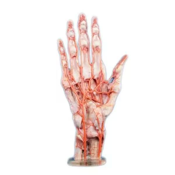
Modern medical institutions face a persistent challenge in spatial visualization. While advanced medical imaging provides detailed internal views, translating two-dimensional screens into tactile surgical realities remains complex. Surgeons and medical students require tactile confirmation to master intricate spatial relationships. High-resolution digital reconstruction combined with advanced printing now addresses this translation issue.
By utilizing continuous transverse sectional images, physical models can be reconstructed to closely match human anatomical proportions. This technological shift is altering how clinical institutions approach medical pedagogy and pre-operative surgical planning.
Bridging the Gap with High-Resolution Datasets
High-fidelity anatomical replication requires a highly accurate data foundation. Standard commercial models often simplify complex structural pathways, which reduces their educational value. To overcome this limitation, DIGIHUMAN utilizes high-resolution digital human datasets as the foundation for anatomical reconstruction. The foundational voxel size of this primary dataset measures 0.0384mm by 0.0384mm by 0.1mm.
Using these ultra-high-resolution datasets, technical systems extract individual voxels from the original sectional data. This process allows for precise surface and internal texture mapping. To verify anatomical accuracy, formalin-fixed human specimens serve as the direct reference during production. This verification process helps the physical model closely replicate the visual structures of human anatomy. The models aim to accurately represent major anatomical structures, including nerves, vessels, and bones.
Why Physical Anatomical Models Improve Spatial Understanding
Transitioning from textbook diagrams to live patients presents a major learning curve. Two-dimensional imaging provides critical diagnostic data but flattens complex anatomical topography. Virtual three-dimensional renderings improve this process by allowing users to rotate and zoom on digital screens. Yet, a screen still lacks true physical depth.
Physical models complete this critical educational progression by transforming digital concepts into tangible reality. Grasping a life-sized model allows learners to better understand the relative scale and spatial relationships of anatomical structures. This direct tactile interaction clarifies orientation within the body cavity. Users can trace the exact path of a nerve around a bone or physically gauge the depth of vascular networks. Holding these accurate structures clarifies how adjacent structures physically interact. This hands-on contact builds the profound spatial relationships necessary for clinical confidence.
Multi-Material Specifications and Printing Capabilities
Translating high-resolution data into a physical model requires specialized fabrication. Standard single-material printing may have limitations when representing different tissue characteristics. To address this, advanced printing systems utilize 12 distinct material channels. This specialized setup integrates high-speed multi-channel inkjet printing with light-curing technology.
The 12 independent channels allow different resins to mix and cure simultaneously. This enables several unique printing modes:
Ⅰ. Single Hardness Printing: Perfect for replicating dense bony structures with rigid, impact-resistant materials.
Ⅱ. Soft and Hard Combinations: Allowing flexible arteries and soft muscle tissue to connect seamlessly with hard skeletal surfaces.
Ⅲ. Transparent Packaging: Enclosing delicate internal pathways inside a clear outer resin block, allowing students to visualize deep structures in three dimensions.
By employing environmentally friendly resin materials, this process produces models with physical characteristics designed to support hands-on anatomical learning. Students and clinicians can gain hands-on experience with anatomical structures through physical model interaction.
Integrating 3D Printed Anatomical Models into Surgical Curriculums
For medical programs and teaching hospitals, integrating 3d printed anatomical models into clinical training protocols improves procedural comprehension. Traditional cadaveric dissection, while highly valuable, presents high recurring costs, regulatory burdens, and limited availability. High-precision replicas provide a repeatable alternative that students can handle indefinitely.
Consider complex structures such as the human hand. The hand features a dense web of overlapping blood vessels, nerves, and small muscle groups. Using a multi-material hand model, students can study the exact depth of the flexor tendons relative to the median nerve. Because the model uses flexible resins for soft tissue, students can manipulate the joints to observe how different muscle groups stretch and interact. This tactile training develops the muscle memory and spatial awareness needed before entering a physical operating room.
Enhancing Pre-Operative Preparation for Complex Surgeries
Beyond standard classroom instruction, multi-material replicas serve as vital assets in pre-operative surgical planning. In complex clinical cases, such as deep facial reconstructions or intricate cranial procedures, surgeons need more than digital imagery. They must rehearse tactile approaches.
By printing a 1:1 scale replica of a patient’s specific anatomical structure, surgical teams can identify potential complications prior to making the first incision. They can test access angles, determine appropriate hardware sizes, and align interdisciplinary teams. This physical rehearsal reduces overall operating times, limits anesthesia exposure, and lowers the risk of structural damage. Having a physical replica available in the operating suite also acts as a visual and tactile reference for the entire surgical team during active procedures.
Combining Digital Anatomy with Physical Replicas
Physical models can provide additional educational value when integrated into a broader digital learning environment. Rather than replacing digital tools, physical models complement systems like the DIGIHUMAN Virtual Anatomy Table.
Students can first perform virtual dissections on high-definition 4K screens to study systemic relationships. They can toggle muscular layers, trace arterial networks, and view sectional planes. Once they understand the digital layout, they can transition to physical, multi-material models to practice tactile identification and surgical hand positioning. This multi-dimensional learning loop links virtual visualization with physical dexterity, preparing students for real-world clinical environments with greater efficiency and confidence.